
Osteodystrophy is a disorder of bone metabolism that results in weakened, poorly mineralized bone tissue, often seen in chronic kidney disease, vitamin D deficiency, or hormonal imbalance. Catching it early relies on precise bone mineral density (BMD) testing and biochemical markers, while prevention mixes nutrition, medication, and lifestyle tweaks.
Why Early Detection Saves Bones
Studies from the National Institute for Health and Care Excellence show that patients diagnosed with osteodystrophy after a fracture have a 30% higher mortality risk than those identified through routine screening. The window between subtle biochemical change and a visible fracture can be as short as 12 months, making timely osteodystrophy detection a lifesaver.
Key Risk Factors to Watch
Understanding what pushes bone health over the edge helps target the right people. The biggest culprits are:
- Chronic kidney disease (CKD) - impaired phosphate excretion and reduced activation of vitamin D create a toxic mineral environment.
- Vitamin D deficiency - low 25‑OH‑vitamin D cuts calcium absorption, driving secondary hyperparathyroidism.
- Elevated parathyroid hormone (PTH) - excess PTH leaches calcium from bone.
- Low serum calcium - signals inadequate mineral supply.
- Sedentary lifestyle and smoking - reduce mechanical loading on bone.
These factors often overlap. For example, CKD patients typically show low vitamin D, high PTH, and abnormal serum calcium, creating a perfect storm for osteodystrophy.
The Diagnostic Toolbox
Doctors have three main imaging options and a suite of blood tests. Choosing the right combination depends on cost, radiation exposure, and the clinical picture.
| Modality | Radiation Dose | Typical Cost (GBP) | Accuracy for BMD |
|---|---|---|---|
| Dual-energy X‑ray absorptiometry (DXA) - the gold‑standard BMD test. | Low | £70‑£120 | High (precision <1%) |
| Quantitative Computed Tomography (QCT) - provides 3‑D volumetric BMD. | Moderate | £150‑£250 | Very high, especially for trabecular bone |
| Magnetic Resonance Imaging (MRI) - assesses bone micro‑architecture without radiation. | None | £300‑£500 | Good for early micro‑damage, but less validated for BMD |
For most primary‑care settings, DXA remains the first line because of its low dose and established T‑score thresholds. When DXA is inconclusive or the patient has metallic implants, QCT or MRI step in.
Biochemical Markers That Speak Before the Scan
Blood work can flag bone turnover days before a density loss shows up on imaging. The core panel includes:
- Serum calcium - low levels suggest inadequate mineral supply.
- Parathyroid hormone (PTH) - high values indicate secondary hyperparathyroidism.
- Alkaline phosphatase - reflects osteoblastic activity.
- 25‑OH‑vitamin D - the circulating form that should stay above 30ng/mL.
Combining these results with imaging sharpens risk stratification. For instance, a patient with a normal DXA but elevated PTH and low vitamin D may still be on the path to osteodystrophy and needs intervention.
Risk Scoring: From Numbers to Action
The FRAX tool calculates a 10‑year fracture probability using age, sex, BMD, and clinical risk factors. A FRAX score above 20% for major osteoporotic fracture usually triggers preventive treatment, even if the DXA T‑score sits at -1.5.
Clinicians also use Z‑scores (age‑matched) for younger patients; a Z‑score <-2 flags early bone loss that warrants further work‑up.
Prevention Strategies That Work
Once risk is identified, a mix of nutrition, medication, and lifestyle changes can halt or reverse bone deterioration.
- Calcium intake: Aim for 1,000mg per day from dairy, leafy greens, or fortified foods.
- Vitamin D supplementation: 800-1,000IU daily keeps 25‑OH‑vitamin D >30ng/mL; higher doses may be needed for CKD patients.
- Phosphate binders: In CKD, agents like sevelamer reduce serum phosphate, lowering PTH drive.
- Bisphosphonates or denosumab: Reserved for patients with confirmed low BMD; they curb bone resorption.
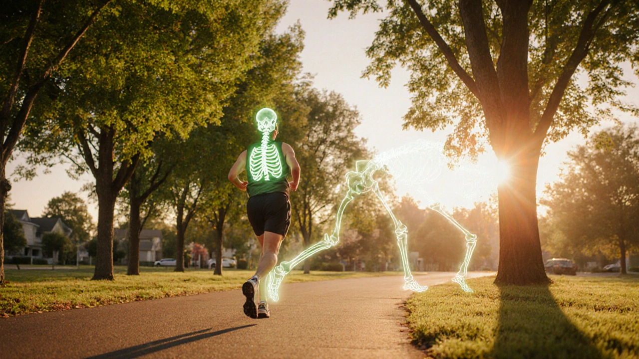
- Weight‑bearing exercise: 30minutes of walking, jogging, or resistance training at least three times a week stimulates osteoblasts.
- Smoking cessation and alcohol moderation: Both improve calcium balance and reduce fracture risk.
Each measure ties back to a core mechanism. Calcium and vitamin D restore the mineral reservoir; phosphate binders correct the CKD‑driven imbalance; exercise provides the mechanical signal that tells bone to stay strong.
Monitoring and Follow‑Up
After initiating prevention, a schedule keeps progress in check:
- Baseline DXA and biochemical panel.
- Repeat DXA at 12‑month intervals for the first two years, then every 2‑3 years if stable.
- Quarterly blood work for CKD patients to adjust phosphate binders and vitamin D doses.
- Annual FRAX calculation to gauge fracture probability trends.
Any upward drift in PTH or a drop of 0.5% in BMD triggers a medication review.
Related Concepts and Next Steps
Osteodystrophy sits alongside other bone‑mineral disorders. Osteomalacia describes a softening of bone due to severe vitamin D deficiency, while Renal osteodystrophy is the CKD‑specific spectrum that includes high‑turnover (osteitis fibrosa) and low‑turnover (adynamic bone disease) subtypes. Understanding these nuances helps clinicians tailor therapy.
Readers hungry for deeper insight can explore:
- “Managing Secondary Hyperparathyroidism in CKD” - a detailed look at PTH pathways.
- “Advanced Imaging for Bone Micro‑Architecture” - a guide to high‑resolution peripheral QCT.
- “Nutritional Strategies for Bone Health” - recipes and supplement timing tips.
Quick Takeaways
- Early osteodystrophy detection saves lives; aim for screening before the first fracture.
- DXA is the first‑line imaging; QCT and MRI fill gaps when DXA is limited.
- Combine BMD with serum calcium, PTH, and vitamin D for a full risk picture.
- FRAX scores >20% merit preventive treatment even with modest BMD loss.
- Calcium, vitamin D, phosphate binders, weight‑bearing exercise, and smoking cessation form the prevention backbone.
Frequently Asked Questions
What is the difference between osteodystrophy and osteoporosis?
Osteodystrophy refers to bone changes caused by systemic disorders like chronic kidney disease or vitamin D deficiency, whereas osteoporosis is mainly an age‑related loss of bone mass without an underlying disease driver. Both increase fracture risk, but treatment pathways differ because osteodystrophy often needs correction of the metabolic disturbance first.
How often should I get a DXA scan if I have early signs of osteodystrophy?
For someone with borderline BMD or abnormal labs, a repeat DXA after 12 months is advisable. If the scan remains stable, extend the interval to every 2‑3 years. Faster follow‑up may be required if PTH spikes or new fractures occur.
Can lifestyle changes alone prevent osteodystrophy?
Lifestyle tweaks-adequate calcium/vitamin D, regular weight‑bearing exercise, and quitting smoking-significantly lower risk, but they often need to be paired with medical therapy when an underlying disorder like CKD is present. Ignoring the metabolic cause can blunt the benefits of lifestyle measures.
What blood tests are most useful for early detection?
The core panel includes serum calcium, phosphate, 25‑OH‑vitamin D, parathyroid hormone (PTH), and alkaline phosphatase. In CKD patients, adding cystatin‑C for renal function helps interpret the mineral results accurately.
Is there a role for MRI in routine screening?
MRI offers detailed pictures of bone micro‑architecture without radiation, but its high cost and limited availability keep it reserved for cases where DXA and QCT are inconclusive or when radiation exposure must be avoided, such as in young adults.
Early detection of bone issues is a game‑changer, especially when you’re juggling CKD or vitamin D deficiency. Getting that DXA scan on schedule can catch subtle drops before a fracture sneaks up. Pair it with a solid supplement plan – calcium, vitamin D, and maybe a phosphate binder if kidneys are struggling. And don’t forget weight‑bearing exercises; even a daily walk up stairs makes a difference. Stick to the routine and you’ll give your skeleton a real fighting chance.
Look, the guide glosses over the real cost burden – you can’t keep throwing money at DXA like it’s a freebie. The system loves to hide the expense while hammering patients with jargon. And this “lifestyle tweak” nonsense? Most folks can’t just quit smoking or start a gym routine overnight. The post needs to be blunt about the socioeconomic barriers.
📊 The numbers don’t lie – serum PTH spikes are a red flag 🚩. Keeping an eye on calcium and phosphate levels weekly can give you a heads‑up before bone loss accelerates. Also, don’t underestimate the power of Mediterranean diet; it’s packed with bone‑friendly nutrients. 🌿💪
From an analytical standpoint, the guide cherry‑picks studies that support its narrative while ignoring the heterogeneity of CKD cohorts. The presented cost figures for DXA ignore regional pricing variance and insurance reimbursements, which can inflate perceived affordability. Moreover, the omission of renal osteodystrophy staging undermines clinical decision‑making. A more rigorous meta‑analysis would reveal that the diagnostic yield of QCT isn’t universally superior. Lastly, the recommendation for quarterly labs is excessive for stable patients and may lead to unnecessary healthcare utilization.
Well, isn’t that just the sweet sound of “simplicity” – as if we can all just pop a vitamin and start lifting barrels. Sure, if you’ve got time to binge‑watch wellness videos and a bank account to match. 🙃 But hey, at least the guide tries to keep it upbeat, right?
I totally get that, bone health matters!
From a coaching perspective, consistency beats intensity. Encourage patients to set a weekly reminder for a short walk or resistance band routine – even 10 minutes counts. Emphasize balanced meals with leafy greens, fortified dairy, and fish oil; the omega‑3s help reduce inflammation that can exacerbate bone turnover. If labs show elevated PTH, a gentle conversation about dietary phosphate sources can be eye‑opening. And always tailor supplement doses to the individual's renal function to avoid hypercalcemia.
Just a quick note – “DXA” should be consistently capitalized, and “vitamin D” needs the proper µg symbol when you mention dosage. Little details keep the guide looking polished.
Let’s make sure everyone feels included in the conversation about bone health. Whether you’re dealing with CKD or just want stronger bones, tailoring the plan to your cultural food preferences can boost adherence. For example, using fortified soy milk instead of dairy works well for many vegans. Keep the language accessible and the recommendations adaptable.
In the grand theater of existence, bone is but a fleeting scaffold upon which we stage our mortal drama. Yet, it whispers of permanence, urging us to contemplate the ephemerality of flesh. Thus, tending to our skeletal foundations becomes an act of reverence toward the fleeting self.
The guide offers a comprehensive overview of osteodystrophy risk factors, diagnostic modalities, and preventive strategies. It correctly emphasizes the importance of routine BMD assessment in high‑risk populations. Moreover, the inclusion of a detailed biochemical panel aligns with current nephrology guidelines. Clinicians should note the recommended interval of 12 months for repeat DXA in borderline cases. Overall, the article serves as a valuable resource for both primary care and specialty providers.
Nice summary, but the piece skirts around the fact that many patients never get access to QCT due to cost constraints.
Wow, yet another “miracle” checklist that ignores the hard truth: most folks can’t afford a dozen scans a year. The guide’s optimism is cute but out of touch with reality. And the casual mention of “lifestyle tweaks” feels like a guilt‑trip for anyone battling chronic illness.
Absolutely! Let’s get those DXA appointments booked!!! 🎉💪 Never underestimate the power of a good vitamin D boost!!!
While the article is well‑intentioned, it suffers from several grammatical inaccuracies. For instance, “the window between subtle biochemical change and a visible fracture can be as short as 12 months” should read “the window … can be as short as twelve months.” Additionally, the list formatting is inconsistent. Correcting these issues will enhance readability. 😊
I appreciate the collaborative spirit of this guide. It brings together nephrology, endocrinology, and nutrition in a cohesive way. Let’s keep the conversation going and share patient success stories. Together we can improve outcomes across the board.
From a philosophical lens, bone health mirrors the resilience of the human spirit. The mineral matrix is a testament to how structure emerges from chaos, much like our own journeys. Embracing preventive care is akin to nurturing the inner fortitude that sustains us. When we honor our bodies, we honor the deeper narrative of existence. Let this guide be a beacon for mindful stewardship of our physical selves.
Minor note: “serum calcium” should be singular when referring to the lab value. Also, avoid using “etc.” in professional writing; list all relevant items explicitly.
Bone health isn’t just a medical issue it’s a cultural one
Let me break this down for anyone who thinks early detection is optional. First, the mortality risk jump isn’t a hypothetical number; it translates to real lives lost when fractures go untreated. Second, the metabolic cascade that begins with a subtle drop in 25‑OH‑vitamin D sets off secondary hyperparathyroidism, which in turn leaches calcium from the very bones we’re trying to protect. Third, the cost of DXA, while modest compared to a hospital admission for a hip fracture, is still a barrier for many, so insurance coverage policies need to be scrutinized and advocated for. Fourth, lifestyle isn’t just a buzzword – weight‑bearing exercise stimulates osteoblast activity, an effect proven in multiple randomized trials. Fifth, smoking cessation isn’t merely about lung health; nicotine directly impairs osteoblast function, worsening bone turnover. Sixth, phosphate binders in CKD aren’t just supplemental; they reduce the mineral load that overwhelms the skeleton. Seventh, consistent monitoring of PTH trends provides a dynamic picture that static calcium levels alone cannot. Eighth, the guide’s suggestion of an annual DXA may be too infrequent for high‑risk patients; some clinicians recommend semi‑annual scans in rapidly progressing disease. Ninth, MRI, while expensive, offers a radiation‑free alternative for younger patients where cumulative exposure matters. Tenth, the biochemical panel should also include fibroblast growth factor‑23 (FGF‑23) in advanced CKD cases to predict vascular calcification risk. Eleventh, patient education materials need to be culturally tailored to improve adherence, especially in diverse populations. Twelfth, interdisciplinary care involving nephrologists, endocrinologists, dietitians, and physiotherapists yields the best outcomes. Thirteenth, we must push for policy changes that fund preventive bone health programs at the community level. Fourteenth, technology like portable bone densitometers could democratize access if funded properly. Fifteenth, ultimately, early detection isn’t just about preventing a fracture; it’s about preserving independence, quality of life, and dignity for our patients.